
Fig. 2. CD40 ligation stimulates the expression of iNOS in IFN-γ-treated BV-2 microglial cells.
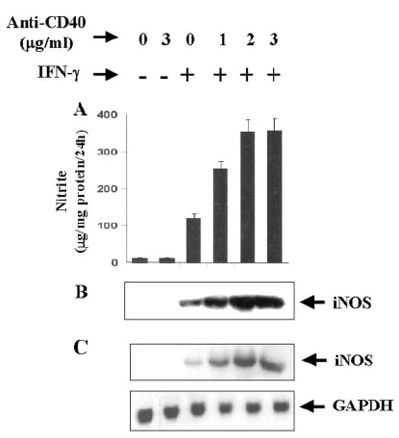
Cells were treated with different concentrations of cross-linking antibodies against CD40 in the presence or absence of IFN-γ (25 units/ml) under serum-free conditions. A, after 24 h, supernatants were used for the nitrite assay as mentioned under “Materials and Methods.” Data are mean ± S.D. of three different experiments. B, cell homogenates were electrophoresed, transferred on nitrocellulose membrane, and immunoblotted with antibodies against mouse macrophage iNOS as described under “Materials and Methods.” C, after 6 h of incubation, cells were taken out directly by adding Ultraspec-II RNA reagent (Biotecx Laboratories Inc.) to the plates for isolation of total RNA, and Northern blot analysis for iNOS mRNA was carried out as described under “Materials and Methods.”