
Fig. 5. Effect of overexpression of CD40 on the expression of iNOS mRNA in BV-2 microglial cells.
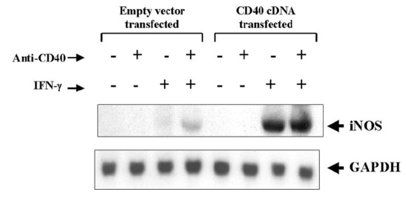
Cells plated at 50–60% confluence in a 100-mm dish were transfected with 0.8 μg of either CD40 cDNA or an empty vector. After 24 h of transfection, cells were stimulated with anti-CD40 (1 μg/ml) in the presence or absence of IFN-γ (10 units/ml) under serum-free conditions. After 6 h of incubation, total RNA was isolated from cells, and Northern blot analysis for iNOS mRNA was carried out.