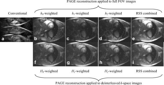
Figure 6.

Images comparing conventional image reconstruction with ghosts (left column) and PAGE reconstructed images (right columns) for ECG triggering segmented multi-echo trueFISP cine acquisition. The top row shows PAGE reconstruction applied to the full FOV image, with individual separated ghost image components (hi weighted) shown in columns 2–4, and the ghost cancelled RSS combined magnitude images shown in right column. The bottom row shows PAGE reconstruction applied to the deinterleaved k-space images, with separate images for each echo time (TE) (Hi weighted) shown in columns 2–4, and the ghost cancelled RSS combined magnitude images shown in right column
