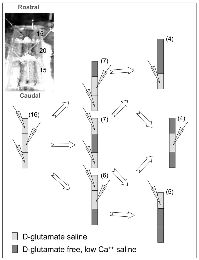
Figure 2.

The experimental preparation was composed of 50 spinal cord segments divided into three chambers as shown (top left, number of segments indicated). Loosely fit suction electrodes were used to record the D-glutamate-induced fictive swimming motor pattern from the ventral nerve roots in the three locations along the preparation. The different experimental manipulations are shown; blocking activity in selected chambers and testing for the effects of depriving short-range or long-range inputs to a selected range of segments whose activity was monitored. (1 mM D-Glutamate, or Glutamate free, low Ca2+ saline were used to induce or block activity respectively)
