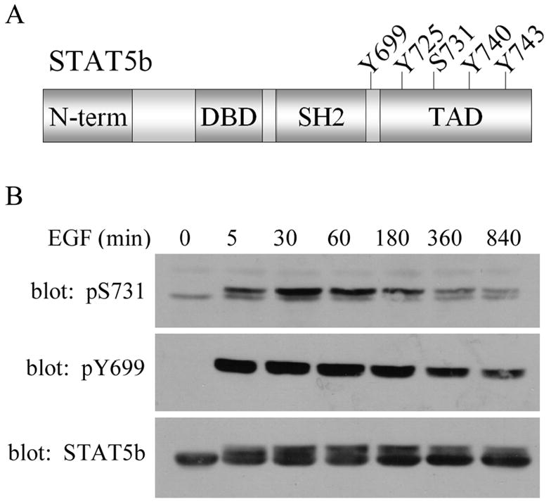
Figure 1. EGF induced STAT5b phosphorylation.

(A) Schematic of STAT5b structure illustrating the conserved domains of the STAT proteins: amino-terminus (N-term), DNA binding domain (DBD), Src homolog domain 2 (SH2), transactivation domain (TAD). The Y and S indicate where tyrosine and serine phosphorylation sites are located. (B) SKBr3 cells stably expressing His-wtSTAT5b were treated with 100ng/mL EGF for the times indicated. Whole cell lysates were analyzed by immunoblotting with antibodies directed against phospho-S731 STAT5b (top), phospho-Y699 STAT5b (middle) and total STAT5b (bottom).
