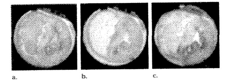
Figure 5.

Three T2-weighted fast SE images without preinversion acquired at (a) 50.5% (b) 100%, (c) 100% hemoglobin saturation. c was acquired 14 minutes after occlusion of the left anterior descending coronary artery. These images demonstrate the BOLD signal intensity modulation as oxygenation levels in the myocardium are adjusted and were also used in the generation of the perfusion images in Figure 4 (third section).
