
Figure 3.
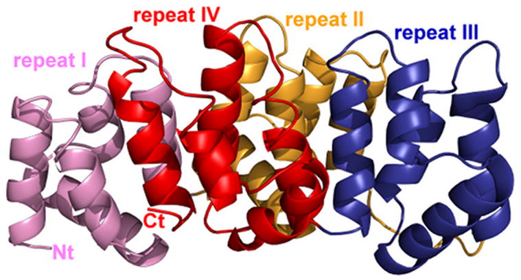
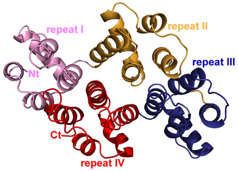
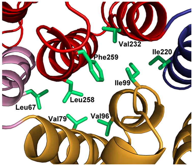
Ribbon diagram of alpha-11 giardin. (a) Side view of the structure with the concave face located at the bottom and the convex face at the top and (b) top view from the convex side. Repeat I is shown in pink, repeat II in gold, repeat III in blue and repeat IV in red. (c) bottom view from the concave side showing the hydrophobic core formed by residues Leu67, Val79, Val96, Ile99, Ile220, Val232, Leu258 and Phe259 (side chains shown in light green). The N-terminus and C-terminus are labeled as Nt and Ct, respectively. The figure was prepared in the molecular graphics program Pymol [http://pymol.sourceforge.net/].