
Figure 7.
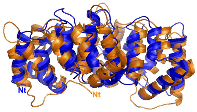
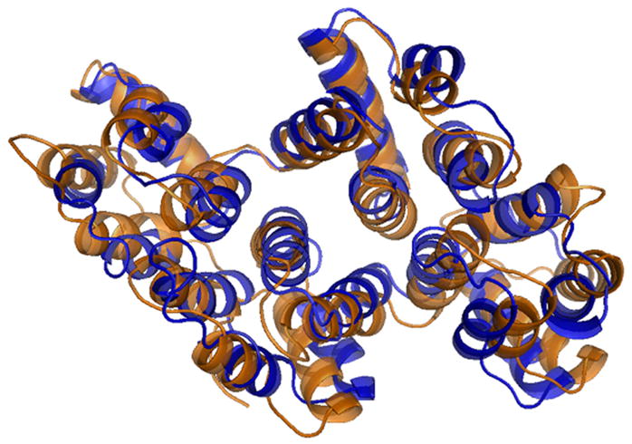
Overlay of alpha-11 giardin (blue) and annexin A4 (orange) crystal structures. (a) Side view of the structure and (b) top view from the convex side. The N-termini (Nt) of alpha-11 giardin and annexin A4 are labeled in their respective colors. Superposition of the alpha-11 giardin and annexin A4 structures was performed in Coot39 and the figures were produced with the molecular graphics program Pymol [http://pymol.sourceforge.net/].