
Figure 8.
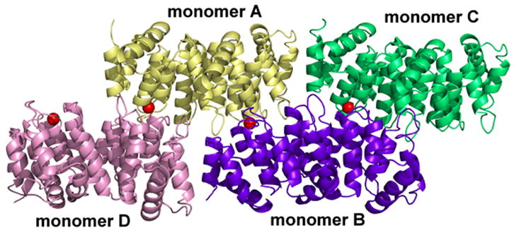
Arrangement of the four monomers in the asymmetric unit of Ca2+-bound alpha-11 giardin. Monomer A is shown in yellow, monomer B in purple, monomer C in green and monomer D in teal. Calcium ions bound to the DE loop of repeat I in each of the four monomers are illustrated as red spheres. The figure was prepared with the molecular graphics program Pymol [http://pymol.sourceforge.net/].