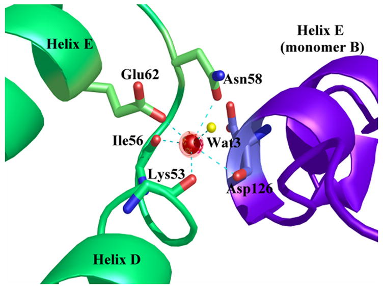
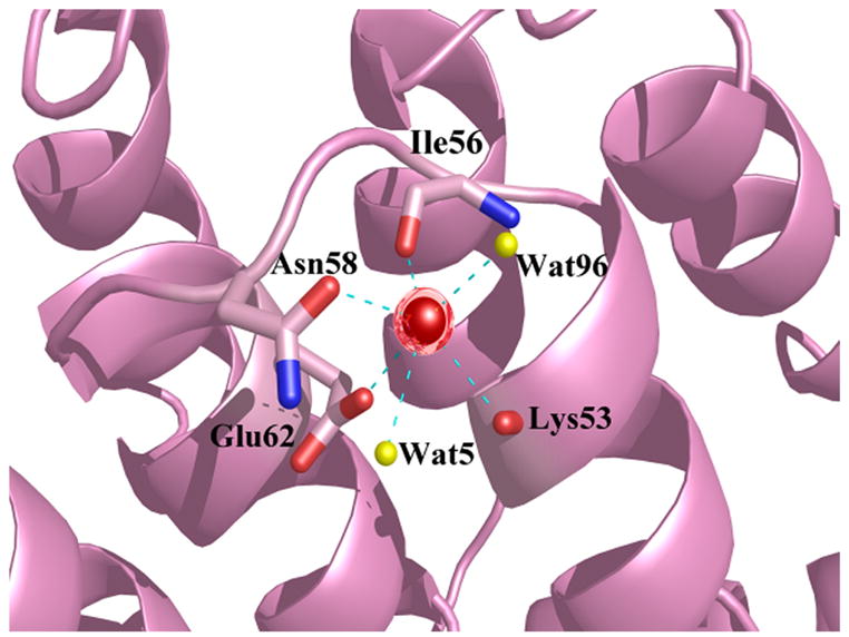
Figure 9.


Coordination of the calcium ion in the DE loop of repeat in Ca2+-bound alpha-11 giardin. Calcium ions are represented as red spheres and water molecules as yellow spheres and the coordination between the calcium ion and oxygen atoms is shown as cyan dotted lines. (a) On the left hand side are the two helices (D and E) from monomer C and on the right hand side is helix E from monomer B (in purple). The coordination distances between the calcium ion in monomer C and the coordinating oxygen atoms (Ca-O) are as follows: Ca-O Lys53 2.83 Å, Ca-O Ile56 2.52 Å, Ca-OD1 Asn58 2.84 Å, Ca-OE1 Glu62 2.71 Å, Ca-O HOH3 2.63 Å and Ca-O Asp126 (monomer B) 2.98 Å. (b) Helices and loops from monomer D are shown in pink. The bond distances between the calcium ion in monomer D and the coordinating oxygen atoms (Ca-O) are as follows: Ca-O Lys53 2.76 Å, Ca-O Ile56 2.75 Å, Ca-OD1 Asn58 2.73 Å, Ca-OE1 Glu62 2.56 Å, Ca-O HOH5 2.89 Å and Ca-O HOH96 2.78 Å. Figures were produced with the programs Pymol [http://pymol.sourceforge.net/] and POVRAY [http://www.povray.org/].
