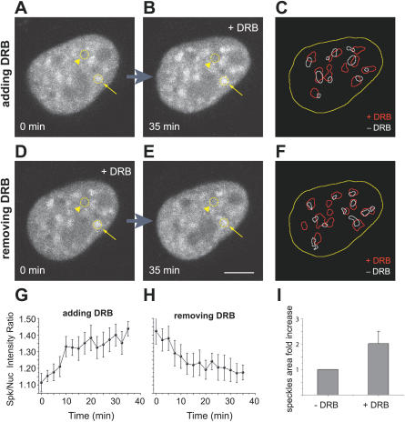
Figure 2. The Transcriptional Inhibitor DRB Induces a Reversible Accumulation of GFP-U2AF65 in Enlarged Nuclear Speckles.
Steady-state distribution of GFP-U2AF65 in the nucleus of a HeLa cell after addition (A,B) or removal (D,E) of DRB.
(A,B) Depict the same cell imaged immediately after addition of DRB (A) and 35 min later (B).
(D,E) Depict the same cell imaged immediately after removal of DRB (D) and 35 min later (E). The arrows point to a nuclear speckle, the arrowheads to the nucleoplasm. Bar indicates 5 μm.
(G,H) Plot of the ratio between fluorescence intensities in the nuclear speckles (n = 6 speckles) and in the nucleoplasm over time after addition (G) or removal (H) of DRB. Error bars represent standard deviations.
(C,F) Depict the threshold segmentations of images in (A,B) and (D,E), revealing the outline of nuclear speckles in the presence (red outlines) and absence (white outlines) of DRB. The nuclear boundary is outlined in yellow.
(I) Quantification of the projected areas in (C,F) reveal an approximate 2-fold increase in speckles size (n = 25 speckles, 4 cells), when transcription is inhibited by DRB.

