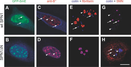
Figure 5. Expression of SPN1ΔN Induces Disassembly of Cajal Bodies and Enlargement of Speckles.
HeLa cells were transfected with either wt snurportin1 (wt SPN1) (A,C,E,G) or the deletion variant SPN1ΔN (B,D,F,H). Cells were imaged for green, red, and blue fluorescence.
(A) Cell cotransfected with GFP-SmE (green) and CFP-wt SPN1 (blue). Arrows point to CBs.
(B) Cell cotransfected with GFP-SmE (green) and CFP-SPN1ΔN (blue). Superimposition of green and blue shows a perfect colocalization of SmE and SPN1ΔN in round and enlarged speckles.
(C) Cell transfected with CFP-wtSPN1 (blue) and immunolabeled with the antibody 4G3 directed against the U2 snRNP B′′ protein (red). Arrows point to CBs and arrowheads to nuclear speckles.
(D) Cell transfected with CFP-SPN1ΔN (blue) and immunolabeled with the antibody 4G3 (red). Superimposition of red and blue images shows colocalization of snRNPs and SPN1ΔN in enlarged speckles (arrowheads).
(E) Cell transfected with GFP-wtSPN1, double-labeled with antibodies directed against coilin (blue) and fibrillarin (red); arrows point to CBs and arrowheads to nucleoli.
(F) Cell transfected with GFP-SPN1ΔN, double-labeled with antibodies directed against coilin (blue) and fibrillarin (red). Note that coilin relocalized from CBs to the nucleolus.
(G) Cell transfected with GFP-wtSPN1, double-labeled with antibodies directed against coilin (blue) and SMN protein (red). Although SMN is detected both in the cytoplasm and in nuclear foci, the cytoplasmic staining is not well-visualized because this image was focused on CBs (arrows), which were in a confocal plane distinct from the cytoplasm. Arrowheads indicate additional minor coilin bodies that are apparently devoid of SMN.
(H) Cell transfected with GFP-SPN1ΔN, double-labeled with antibodies directed against coilin (blue) and SMN (red); arrowheads point to nucleoli that now accumulate coilin and arrows point to nuclear SMN foci. Bar indicates 10 μm.

