Figure 1.
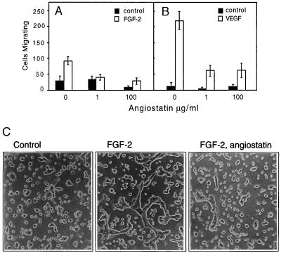
(A and B) Angiostatin attenuates migration of HUVE cells toward FGF-2 (A) and VEGF (B). Cell migration was measured in a Boyden chamber, and the cells migrating through the filter during a 4-h incubation were stained and counted. (C) Angiostatin treatment of murine brain endothelial cells leads to diminished formation of tubes in three-dimensional collagen gels. Murine brain endothelial cells were seeded out on a solidified collagen gel and covered with a second layer of collagen. After 10 h of treatment in the presence or absence of growth factor and angiostatin, the cells were examined for formation of tube-like structures. The cells were treated as indicated with 10% fetal calf serum alone (Left), with 10% fetal calf serum and 5 ng/ml FGF-2 (Center), or pretreated for 72 h with 2.5 μg/ml of angiostatin in Ham’s F-12, 10% fetal bovine serum, followed by inclusion of angiostatin in the collagen gel and stimulation of tube formation by 5 ng/ml FGF-2 (Right). Angiostatin treatment led to 79% decreased formation of tubes, from 305 μm total tube length in the absence (−angiostatin) to 65 μm total tube length in the presence (+) of angiostatin. (×200.)

