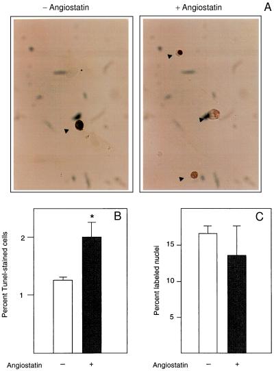
Figure 2.
(A) Angiostatin induces apoptosis but has no effect on DNA synthesis in endothelial cells. BCE cells cultured in the presence of 10% serum on coverslips were kept in the absence (−) and presence (+) of 2.5 μg/ml angiostatin for 3 days, at which point the cells were fixed and processed for TUNEL staining (12), to detect cells undergoing programmed cells death. Cells stained positive in the TUNEL assay are indicated by arrowheads. (B) Number of TUNEL-positive cells in the untreated (open bar) and angiostatin-treated (solid bar) cultures. Values are percentage of positive cells ± SEM for four determinations. ∗ indicates P < 0.05. (C) Number of nuclei incorporating [3H]thymidine in the untreated (open bar) and angiostatin-treated (solid bar) samples. Cells were plated on coverslips and cultured in the presence of 10% newborn calf serum with or without 2.5 μg/ml angiostatin for 3 days. During the last 2 h of culture, 1 μCi/ml [3H]thymidine was present in the culture medium. The cells were washed, fixed, and processed for autoradiographic determination of [3H]thymidine incorporation. Values are percentage of labeled nuclei ± SEM for three determinations.

