
Fig. 6.
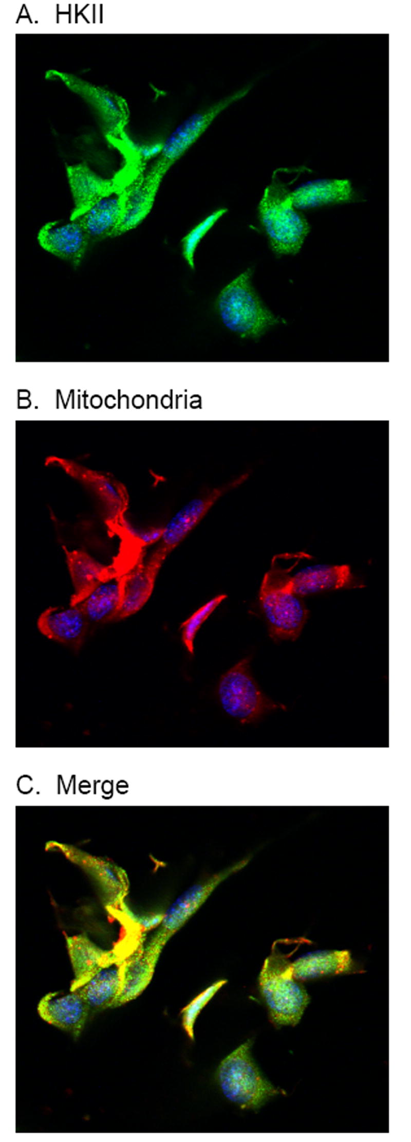
Immunofluorescence imaging of arsenic treated mesangial cells. Cultured mesangial cells were treated with 50 ppb arsenic for 24 hours, then fixed and stained. HKII was with an isoform specific primary and matched fluorescent secondary antibody (A), mitochondria were stained with MitoTracker Deep Red (B), and nuclei counterstained with DAPI (blue). The merged image (C) shows the co-localization of HKII with the mitochondria.