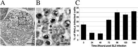
Figure 2. C. caviae Undergo a Full Developmental Cycle in Drosophila SL2 Cells.
(A) Electron micrograph of semi-thin sections (∼70 nm) of Drosophila SL2 cells 45 h post infection with C. caviae. Black square: Area magnified and shown in (B). Bar: 2 μm.
(B) Higher magnification of the bacteria of the C. caviae inclusion shown in (A). Bar: 500 nm.
(C) C. caviae were isolated from Drosophila SL2 cells 3, 24, 48, 72, 96, 120, and 144 h post infection and were used to infect HeLa cells for 24 h. The percentage of HeLa cells that present an inclusion is shown.

