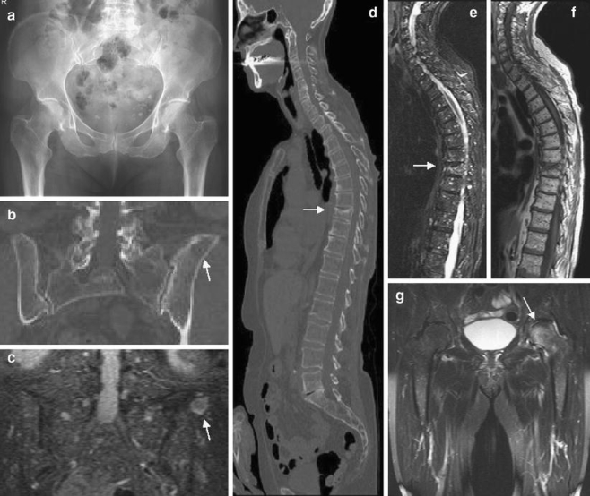
Fig. 3.

A 70-year-old man with multiple myeloma. a The radiograph of the pelvis is inconspicuous. b Coronal MS-CT reconstruction of the pelvis in a bone window setting reveals a large area of destruction within the left iliac bone (arrow). c) STIR-WB-MRI confirms focal tumor manifestation within the iliac bone (arrow) and reveals multiple small nodular infiltrations within the sacral bone and pelvis. d MS-CT of the spine shows a compression fracture of Th9. e, f T1-weighted SE- and STIR imaging of the spine reveals diffuse myeloma infiltration of the spine. g Coronal STIR sequences of the pelvis show additional focal infiltration of the left femoral head missed on radiography and MS-CT
