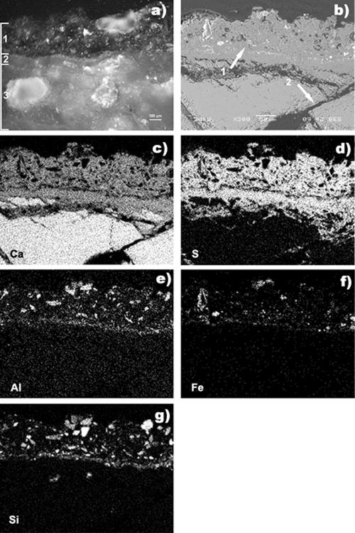
FIG. 2.
Polished cross section of sample 1 surface before cleaning. (a) Optical microscopy. Layer 1, black crust layer; layer 2, yellow-ochre layer (patina); layer 3, marble substrate. (b) SEM observation (back-scattered image). Arrow 1, black crust; arrow 2, microfractured substrate. (c to g) EDX analysis. (c) Calcium map. (d) Sulfur map. (e) Aluminum map. (f) Iron map. (g) Silicon map.

