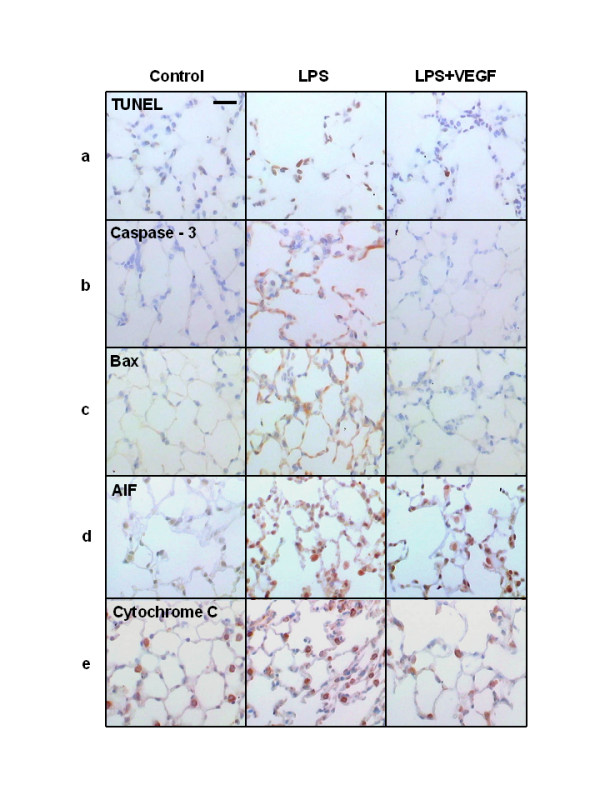
Figure 2.

Representative appearances of lung tissue specimen after TUNEL and immunohistochemical staining. a) In the LPS group, characteristic chromatin condensation in the nuclei of TUNEL-positive epithelial and endothelial cells were observed, which were decreased in the LPS+VEGF group. b-e) Caspase-3 (b), Bax (c), AIF (d) and cytochrome C (e) immunostaining were present in epithelial and endothelial cells, but not in macrophages and neutrophils. In the LPS+VEGF group, TUNEL, caspase-3, Bax, AIF and cytochrome C positive cells were decreased compared with the LPS group. Scale bar = 50 μm.
