
Figure 2.
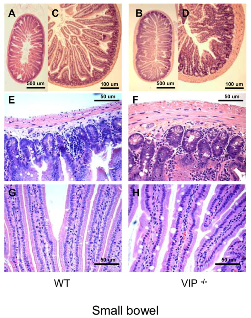
Microscopic structure of the proximal jejunum in WT and VIP−/− mice. Small bowel sections were stained using standard H&E staining. Pictures at low (x2.5, x10, upper panel) and higher magnification (x20, x40; mid and lower panels, respectively) revealed an overall increase in thickness of muscularis mucosa in VIP KO (B, D and F) vs. controls (A, C and E), as well as higher numbers of mucus-forming cells (G vs. E).