
Figure 4.
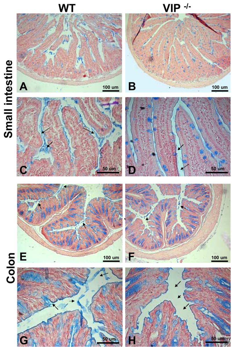
Alcian blue staining to visualize mucus deposits on sections in duodenum (A–D) and distal colon (E–H) from WT (left panel) and VIP−/− mice (right panels). As expected from H&E staining shown in figures 2 & 3, Alcian blue staining revealed a significant difference in mucus-positive cells in the small intestine, but no change in colon. However, sections from VIP−/− showed a dramatic reduction in the amount of mucus released in the lumen in both small (A, C vs B, D), and large (E, G vs F, H) bowels.