
Figure 5.
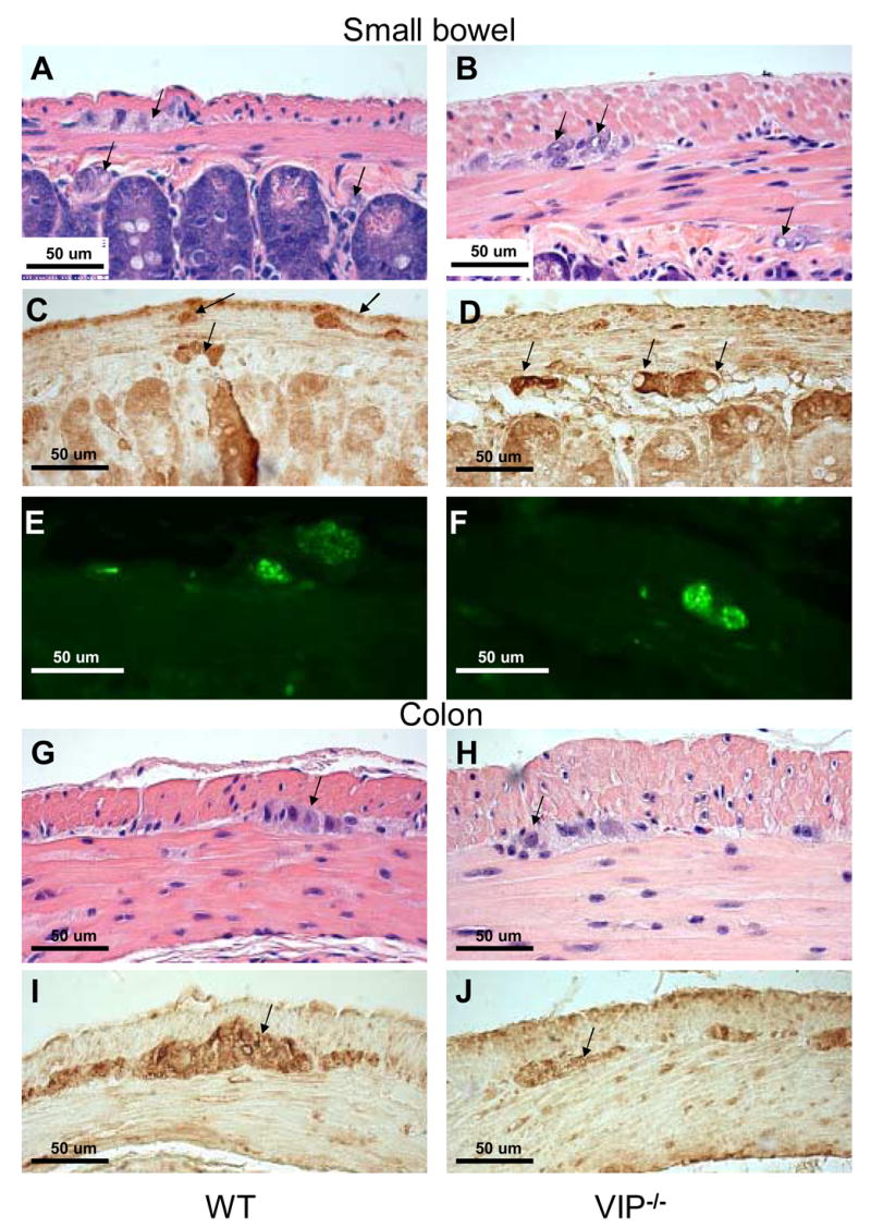
Microscopic structure of myenteric plexus in duodenum and distal colon of WT and VIP−/− mice. H&E (A, B, G and H) and S100β (C–D & I–J) and pan-neurofilaments (NF) (E, F) stainings are shown in WT and VIP KO mice (left and right panels, respectively). Structural differences were observed in duodenum of VIP deficient mice when compared to WT controls. Plexuses from KO mice showed enlarged unstained patches (A vs B) that were non-immunoreactive with S100β or NF antibodies used to specifically reveal Schwann cells and axons (C vs. D & E vs. F, respectively). However, immunofluorescence emitted by the pan-NF antibody coupled with green fluorescent dye-labeled conjugates (E vs. F) revealed larger area signals within the external plexus of the KO mice than WT controls, suggesting the presence of bigger axons within the myenteric plexus of VIP-deficient mice. Conversely, no obvious differences in plexus structures of the large bowel (G–J) were using these techniques (pan-NF not shown).