Figure 2.
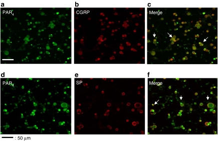
Immunohistochemical detection of PAR4 and colocalization with CGRP and SP in cultured DRG neurons. Culture dishes were incubated in the presence of anti-PAR4 antibody (a, d), in addition to an incubation with anti-CGRP antibody (b), or an SP antibody (e). Panels c and f represent the merging of images from panels a and b, and panels d and e, respectively. Scale bar represents 115 μm.

