
Figure 2. [3H]DAA1106 binding corresponds to microglia in rats with TBI.
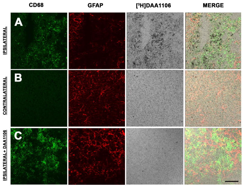
A–B, Combined [3H]DAA1106 autoradiography (black grains) and immunostaining for activated microglia (CD68, green) and astrocytes (GFAP, red) was performed in cortical brain tissue in rats with CCI on the ipsilateral side at the site of contusion (A) and on the contralateral side (B). On the ipsilateral side (A), [3H]DAA1106 Specific binding overlapped with CD68 labeled microglia but not with GFAP immunostained astrocytes (vertical panel - Merge). On the contralateral side (B), minimal CD68 and [3H]DAA1106 binding was seen.
C, [3H]DAA1106 autoradiographic binding was confirmed to be specific as it was displaced by unlabeled DAA1106 in adjacent sections on the ipsilateral side. Scale bar indicates 50 μm.