
Figure 4. [3H]DAA1106 binding is higher in regions with neuronal loss.
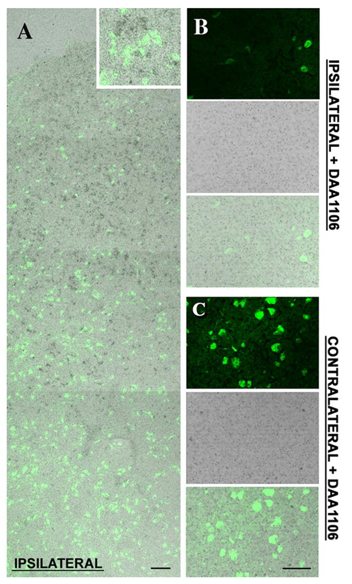
A, Montage image of combined NeuN staining (green) and [3H]DAA1106 autoradiography (Black grains) in cortical brain section obtained from rats with CCI. NeuN staining did not overlap with [3H]DAA1106 binding (inset). NeuN staining progressively decreases as the site of the lesion is approached indicating neuronal loss. Scale bar indicates 50 μm.
B & C, [3H]DAA1106 autoradiographic binding was confirmed to be specific on the ipsilateral side (B) and on the contralateral side (C) as it was displaced by unlabeled DAA1106. Scale bar indicates 50 μm.