
Figure 1.
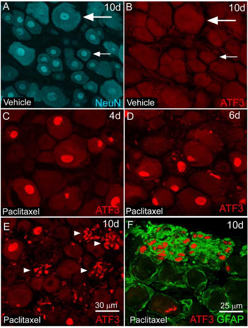
Activating transcription factor 3 immunoreactivity (ATF3-IR) is increased in neurons and non-neuronal satellite cells in L4 dorsal root ganglia (DRG) of rats following intravenous paclitaxel administration. L4 DRG section from paclitaxel (C-F) and vehicle (cremephor/ethanol) treated rats (A,B) were examined immunohistochemically with antibodies against activating transcription factor (ATF3, red) a marker of cellular injury/regeneration at several time points following administration of paclitaxel. Sensory neurons in DRG were identified immunohistochemically with antibodies against NeuN (NeuN, blue) a neuronal nuclei marker (A) that labels both small (small arrow) and large (large arrow) diameter sensory neurons. Increased ATF3 immunoreactivity (IR) was observed in neuronal nuclei at day 4 (C) day 6 (D) and to a lesser extent at day 10 post paclitaxel administration (E). At day 10, there was also an increase in clusters of ATF3-IR satellite cells (E, arrowhead) within the DRG of paclitaxel-treated rats as these cells coexpressed the intermediate filament glial fibrillary acidic protein (F, GFAP, green). No ATF3 expression was observed in L4 DRG of vehicle-treated rats at day 10 (B) or other time points examined. Scale bar: A-E 30 μm. F: 25 μm.