
Figure 6.
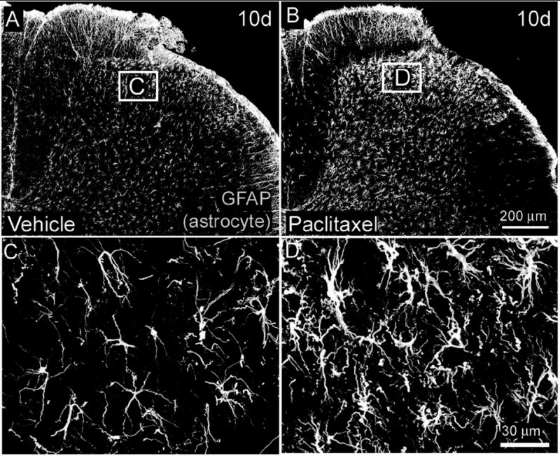
Glial fibrillary acidic protein immunoreactivity (GFAP-IR) is increased in astrocytes within the L4 spinal cord of paclitaxel-treated rats. Beginning six and ten days (B) following intravenous administration of paclitaxel an increase in GFAP-IR is present in astrocytes throughout the dorsal horn (laminae I-VI) of the L4 spinal cord of paclitaxel-treated rats (B) compared to vehicle (cremophor/ethanol) treated rats (A). Higher magnification demonstrates that astrocytes in paclitaxel treated rats posses a more hypertrophic appearance typical of activated astrocytes (D) compared to vehicle-treated rats (C). Scale bar A, B = 200μm; C, D = 30μm.