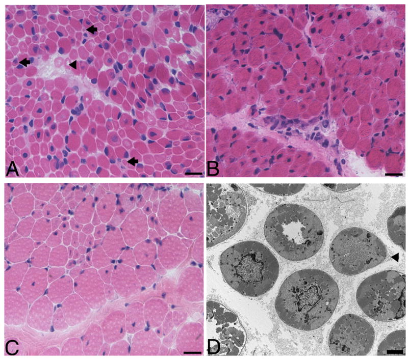
Figure 1.

(A) H&E stained frozen section of the quadriceps biopsy performed at 6 days of age from patient 447-1, who has an MTM1 mutation that truncates myotubularin shows scattered centrally nucleated hypotrophic myofibers with the characteristic appearance of myotubes (arrowhead). Note the presence of many extremely small myofibers (arrows). (B) H&E stained frozen sections from patient 171-1a with R69C missense mutation and had a quadriceps biopsy at 8 days. Note that there are no extremely small myofibers like those depicted in A. (C) The contralateral quadriceps was biopsied in patient 171-1b at 3.3 years and it shows a population of considerably larger fibers. (D) Electron micrographs show small rounded myofibers with central nuclei and glycogen, which was qualitatively similar whether a patient had an MTM1 missense mutation or truncation/deletion mutation. Note the redundant basal lamina suggestive of atrophy (arrowhead). The bar is 20 μm in A, B and C and 2 μm in D.
