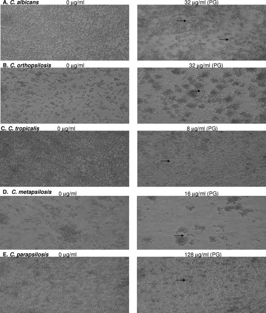
FIG. 3.
Light microscopy of representative biofilms of each of the Candida sp. tested which displayed PG in biofilm MIC tests. Biofilms in the drug-free control well and biofilms displaying PG in the presence of high CAS concentrations after 48 h incubation are shown. (A) C. albicans; (B) C. orthopsilosis; (C) C. tropicalis; (D) C. metapsilosis; (E) C. parapsilosis. Arrows point to the “globose” cells frequently observed in wells displaying PG.

