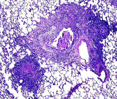
FIG. 1.
Lungs from an M. tuberculosis-infected guinea pig showing primary and secondary lung lesions. A residual primary lung lesion with dystrophic calcification, which is surrounded by mixed inflammation representing secondary lesions resulting from hematogenous dissemination and inflammation, is shown. Magnification, ×100 (H&E stain).

