
FIG. 1.
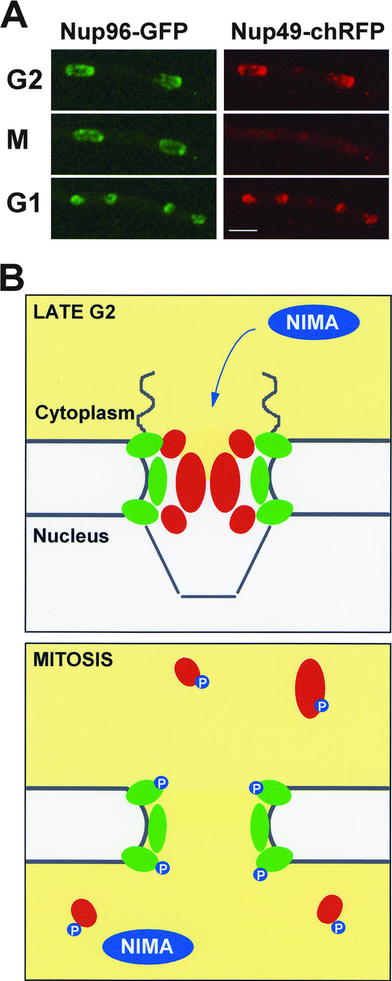
Partial NPC disassembly during mitosis in A. nidulans. (A) Time-lapse images of Nup96 labeled with GFP and Nup49 labeled with the mCherry variant of red fluorescent protein (chRFP) in the same cell (43). During G2, both Nups locate to NPCs at the nuclear periphery. However, as the cell enters mitosis, Nup49 disperses throughout the cell, while Nup96 remains at the nuclear periphery. As cells exit mitosis, Nup49 reassociates with the nuclear periphery of the G1 nuclei (see movie S1 in the supplemental material). Bar, ∼4 μm. (B) Schematic model of an A. nidulans NPC during late G2 and mitosis. In late G2, FG-repeat Nups (red) occupy the central channel of the NPC, restricting passive diffusion and helping to facilitate active transport. Accumulation and activation of the NIMA kinase during G2 trigger the dispersal of central channel Nups, while core Nups (green) remain associated with the nuclear envelope. Putative phosphorylation of Nups by NIMA is indicated. Opening of the NPC central channel compromises active transport and also allows passive diffusion through the NPC. This allows equilibration between the cytoplasm and the nucleus and thus tubulin to gain access to nuclei specifically during mitosis.