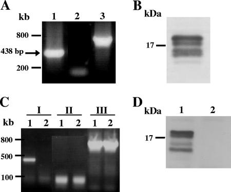
FIG. 3.
mRNA and protein expression of EhLimA. (A) An RT-PCR analysis of total RNA using primers specific for EhLimA results in amplification of a band of the expected size of 438 bp (lane 1). No amplification is attained when the reverse transcription reaction is performed without reverse transcriptase (lane 2). Amplification of actin is shown in lane 3. (B) Detection of EhLimA in a Western blot analysis of total cell lysates using purified anti-EhLimA antibodies reveals four bands around 16 kDa. (C) An RT-PCR analysis of total RNA of G3 transfected trophozoites in which EhLimA is transcriptionally silenced (lane 2) and of nontransfected G3 cells expressing EhLimA (lane 1). Amplification of EhLimA (lanes I) is attained only with the nontransfected G3 cells but not with the EhLimA-silenced cells. No amplification is attained when the reverse transcription reaction is performed without reverse transcriptase (lanes II). Amplification of actin (lanes III) is attained with both the nontransfected G3 cells and with the EhLimA-silenced cells. (D) Western blot analysis of total cell lysates of G3 transfected trophozoites in which EhLimA is transcriptionally silenced. The anti-EhLimA antibodies do not detect any protein in these cells (lane 2), whereas the protein is detected in the nontransfected G3 cells expressing EhLimA (lane 1).

