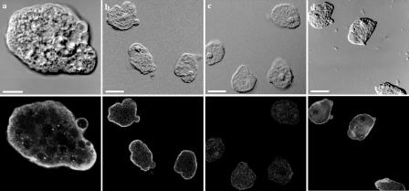
FIG. 4.
Immunofluorescence localization of EhLimA in whole parasites. Trophozoites were fixed and stained for immunofluorescence with anti-EhLimA antibodies followed by fluorescein isothiocyanate-conjugated anti-rabbit secondary antibodies and viewed under a confocal microscope. The differential interference contrast images are shown on the top, and the corresponding fluorescence images are shown on the bottom. EhLimA localizes mainly to the plasma membrane of cells (a and b). No staining was detected in the EhLimA-silenced G3 amoeba cells (c). Staining cells with phalloidin-TRITC reveals and enrichment of actin in the cell cortex and in cellular protrusions (d). Bars, 10 μm in panel a and 20 μm in panels b, c, and d.

