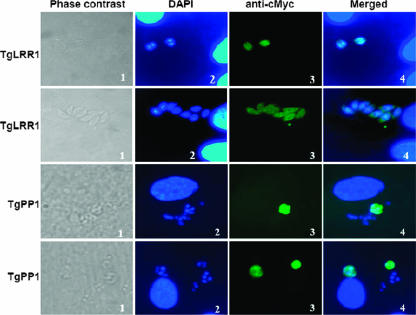
FIG. 8.
Localization of T. gondii tachyzoites TgLRR1 and TgPP1 using transient transfection assays. Two independent sets of data for TgLRR1-cMyc are shown (two upper sets, magnification ×100). Panel 1, phase contrast; panel 2, DAPI staining; panel 3, IFA using anti-cMyc; panel 4, merged DAPI and IFA. Two independent sets of data for TgPP1-cMyc are shown (two lower sets, magnification ×65). Panel 1, phase contrast; panel 2, DAPI staining; panel 3, IFA using anti-cMyc; panel 4, merged DAPI and IFA. Note that TgLRR1 and TgPP1 signals are predominantly seen in the parasite's cytoplasm, whereas a weak but consistent nuclear fluorescence can be observed.

