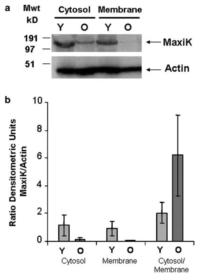
Figure 2.

Subcellular fractionation and Western blotting to localize expression of the MaxiK channel. (a) A typical Western blot where expression of MaxiK in the cytosolic and membrane fractions of corpora is analysed in young (Y) and old (O) animals. Actin is used to normalize loadings. (b) Densitometric analysis of protein expression. Four animals, two young and two old were analysed by Western blot in duplicate. Expression of MaxiK and actin protein was determined in samples by densitometry. Expression of MaxiK was normalized by comparison with actin. The columns represent the mean of four analysis, and the error bars the average deviation from the mean. The expression of MaxiK is shown in the cytosol and membrane fractions, and also as a ratio of the amount in cytosol to membrane fractions in young (Y) and old (O) animals.
