
Figure 3.
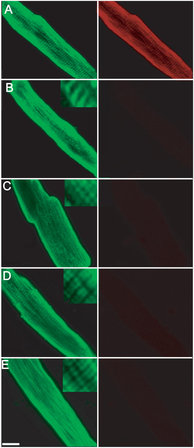
Representative immunofluorescence projection images of myocytes expressing ssTnI substitution mutants 6 days after gene transfer. Myocytes were immunostained with MAB1691 (left panel), which recognizes all troponin I isoforms and a FITC-conjugated secondary antibody, while cTnI-specific staining was detected with TI-1 antibody (right panel) and a Texas-Red-conjugated secondary antibody. Immunostaining of cTnI (TI-1 Ab) disappears in myocytes expressing ssTnI and ssTnI substitution mutants (right panels of B–E). The striated pattern of TnI staining using the non-isoform-specific MAB1691 indicates the ssTnI mutants are incorporated into the myofilaments. A. Control myocytes, B. ssTnIR125Q, C. ssTnIH132A, D. ssTnIV134E, and E ssTnIQAE. A similar pattern of staining was obtained in myocytes expressing ssTnIQAETAT (results not shown).