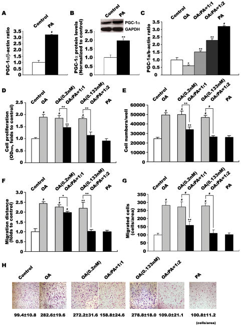
Figure 4. Changes of PGC-1α expression and VSMC proliferation/migration in response to increased proportion of PA in fatty acid mixtures.
VSMCs were incubated with various FFAs for 24 h before analysis. Effects of PA on PGC-1α expression was determined by quantitative PCR (A) and western blot analysis (B). Effects of increased PA on PGC-1α expression were determined by quantitative PCR (C). Effects of increased PA on OA-induced VSMC proliferation were determined by MTT assay (D) and cell counting (E). Effects of increased PA on OA-induced VSMC migration were determined by wound healing (F) and transwell analysis (G,H). Data in VSMC proliferation and migration detections represent the means±SEM of 18 determinants from 3 independently prepared samples each with 6 measurements. Data in PGC-1α mRNA level detections are expressed as means±SEM of five different experiments normalized to β-actin levels. Data in PGC-1α protein level detections are expressed as means±SEM of four different experiments normalized to GAPDH levels. *P<0.05, **P<0.01, #P<0.001 vs. control

