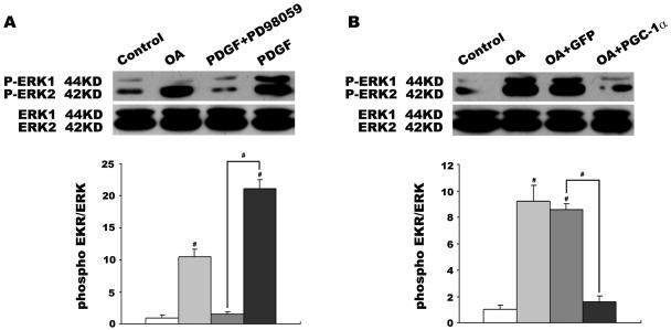
Figure 5. Effects of PGC-1α on OA-induced p-ERK activity in VSMCs.
PDGF-BB and pharmacological ERK-MAPK inhibitor PD98059 were chosen as positive and negative controls of ERK phosphorylation, respectively. VSMCs were made quiescent by serum-starvation for 24 h and then stimulated with PDGF-BB (100 ng/ml) for 30 min, PD98059 (50 uM) for 1 h before stimulation with PDGF-BB (100 ng/mL), or 0.4 mmol/L OA for 2 h. Proteins extracted following these treatments and phosphorylation of ERK was determined by western blot (A). VSMCs were also treated with 48 h adenovirus infection and then incubated with 0.4 mmol/L OA for 24 h. Phosphorylation of ERK was also analyzed with elevated PGC-1α level by western blot (B). Data were shown as the ratio of p-ERK/total ERK. The ratios of control were designated as 1.0. Individual data in this chart represent the mean±SEM of 4 determinants. #P<0.001 vs. control or positive/negative group.

