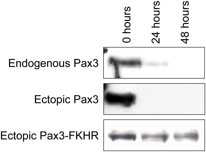
Figure 1.
Expression of Pax3 and Pax3-FKHR during myogenic differentiation. Mouse primary myoblasts (top panel) and mouse primary myoblasts stably expressing either Pax3 (middle panel) or Pax3-FKHR (bottom panel) were induced to differentiate as described in the Materials and Methods. Undifferentiated primary myoblasts (0 hours) and myoblasts from each time point of differentiation (24 and 48 hours) were lysed, equal amounts (50μg) of total cell extracts were separated by 10% SDS-PAGE, the equal loading of protein was confirmed by Fast Green staining of the membrane (data not shown), and the presence of endogenous Pax3, ectopic Pax3, or ectopic Pax3-FKHR was determined by Western blot analysis using a monospecific affinity-purified anti-Pax3 antibody, as described in the Materials and Methods.

