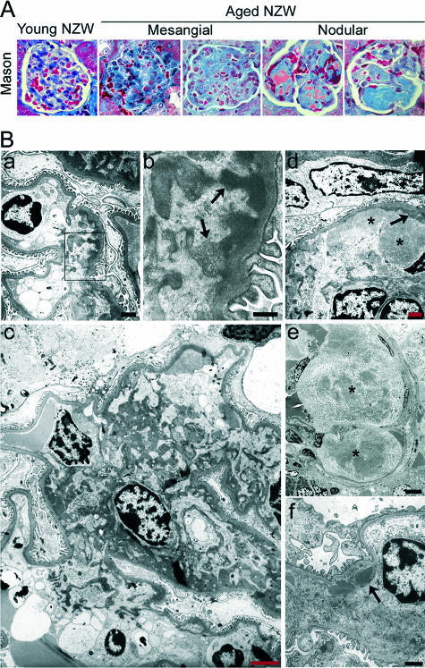
Figure 1.
NZW mice undergo age-dependent glomerulonephritis. A: Mason trichromic staining of glomerular sections representing the indicated mice and glomerulonephritis (n = 60). In this and following figures, young is <6 months and aged is >7 months. B: Glomerular ultrastructural pathological findings in aged NZW mice (n = 5). a and b: Electron-dense homogenous or vacuolated materials aligned on lamina densa of (para)mesangial GBM are denoted by arrows in b, which represents the boxed region in a analyzed at higher magnification. c: Electron-dense homogeneous material filling mesangium and paramesangium. d and e: Electron-dense vacuolated material occupying paramesangium (d) and capillary space (e). f: Electron-dense material associated with lamina rara of (para)mesangial GBM. In d–f, arrows and asterisks denote electron-dense homogenous and vacuolated material, respectively. Illustrated are selected images from NZW mice of 5, 10, 13, 9, and 9 months (left to right) (A) and 8 to 9 months (B). Scale bars: 1 μm (a, d, and f); 0.5 μm (b); 2 μm (c); 7 μm (e). Original magnifications, ×400 (A).

