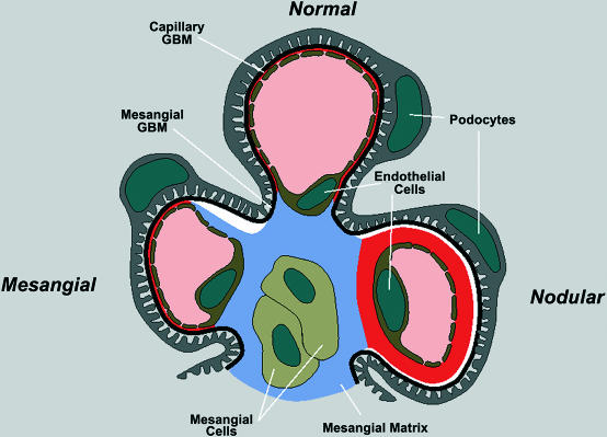
Figure 7.
Model for type IV collagen distribution in normal glomeruli and in GPBP-mediated glomerulonephritis. The clover-like structure represents a section of a renal glomerulus in which type IV collagen is depicted in an unaffected capillary (normal) or in capillary undergoing the indicated GPBP-mediated glomerulonephritis. In black, the α3.α4.α5(IV) collagen (epithelial GBM component). In red, membrane-organized α1.α1.α2(IV) collagen (endothelial GBM component). In blue, mesh-organized α1.α1.α2(IV) collagen (mesangial component). In white, virtual spaces resulting from defective fusion of epithelial and endothelial GBM components that support GPBP accumulation and IgA deposits on orphan epithelial wall. In mesangial glomerulonephritis, the mesangial component progressively replaces the endothelial component whereas in nodular lesions the endothelial component progressively substitutes mesangial component. In advanced glomerulonephritis, a component can entirely substitute the other. In both NZW and Tg-hGPBP mice, there were animals, glomeruli, and even capillaries displaying both types of lesion.

