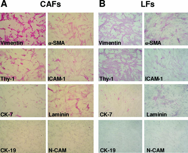
Figure 2.
Representative images of cultured CAFs (A) and LFs (B) stained for vimentin, α-SMA, Thy-1, ICAM-1, cytokeratin (CK)-7, laminin, CK-19, and neural cell adhesion molecule (NCAM), indicating that the myofibroblastic phenotype of CAFs and LFs was stable in vitro (original magnification, ×400; positive staining, red).

