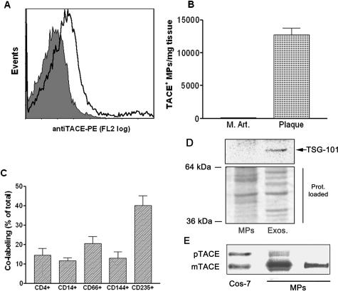
Figure 1.
MPs isolated from atherosclerotic human plaque, which contain the mature form of TACE/ADAM17, are of diverse cellular origin and do not contain exosomes: Analysis of TACE/ADAM17 on MPs isolated from human atherosclerotic plaque and human internal mammary arteries. A: Expression of TACE/ADAM17 on MPs from plaque homogenates. This graph is representative of the different plaque preparations. The shaded peak corresponds to negative isotype control. B: Levels of TACE/ADAM17+ MPs in human internal mammary arteries (M.Art., n = 3) and atherosclerotic plaque (plaque, n = 25); values are mean ± SEM. C: Co-labeling of TACE/ADAM17+ MPs with various cellular markers (from left to right: lymphocytes, monocytes, granulocytes, endothelial cells, and erythrocytes) (n = 12). Results are expressed as percentage of total TACE/ADAM17+ MPs (mean ± SEM). D: Immunoblotting of the exosomal marker TSG-101 in the post 20,500 × g pellet (left) and corresponding supernatant further centrifuged at 170,000 × g (right). Because protein profiles were different in both fractions (see Ponceau red staining), two times more MP materials (20 μg) than exosomal-like material were loaded. Representative of three different samples analyzed. E: Immunoblotting of TACE/ADAM17 from two different MP preparations containing 2.8 × 10⁶ and 0.1 × 10⁶ Annexin V+ MPs/μl on the middle and right lanes, respectively. The TACE/ADAM17 of MPs in the middle lane is to illustrate that only a highly MP-enriched plaque allows the detection of TACE proform. The right lane is representative of the five MP preparations tested. mTACE and pTACE indicate the positions of the mature and proform of TACE/ADAM17, respectively, validated by the migration of these forms present in COS-7 cells.

