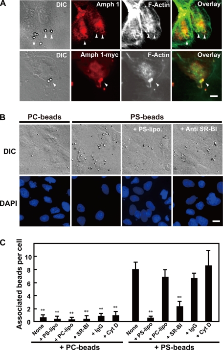
Figure 2.
PS-dependent phagocytosis in Sertoli cells. (A) Amphiphysin 1 accumulates at phagosomes. Ser-W3 cells were transfected with amphiphysin 1-myc. After 24 h of transfection, the transfected cells were incubated with PS beads at 37°C for 90 min. They were fixed, permeabilized with digitonin, and stained with anti-amphiphysin 1 antibodies (mab 3) (top) and anti-myc antibodies (bottom) and Alexa488-phalloidin. Arrowheads indicate the incorporated beads surrounded by amphiphysin 1 and polymerized actin. Bar, 5 μm. (B and C) The association of beads with the Ser-W3 cells is PS and SR-BI dependent. Cells (1 × 10⁴ cells on coverslips) were pretreated with 0.25 mM liposomes containing PS or PC at 37°C for 10 min. Then, cells were incubated in presence of beads coated with PS- or PC-liposomes at 37°C for 180 min. The cells were then washed, fixed, stained with DAPI, and analyzed by phase contrast microscopy. To block the binding of PS receptors to PS beads, cells were pretreated with anti-SR-BI antibodies or rabbit IgG as negative control at 100 μg/ml for 30 min (B). Bar, 10 μm. The number of beads associated with the cells was determined using phase contrast microscopy, and it is presented as the number of the beads per cell. To block the internalization, cells were pretreated with Cyt D at 2 μM for 30 min, and attached beads to the cells were analyzed. One hundred cells in 10 independent fields were counted in each experiment. All results are the mean ± SEM from three experiments. Statistical significance was determined by Student's t tests (**p < 0.01) (C).

