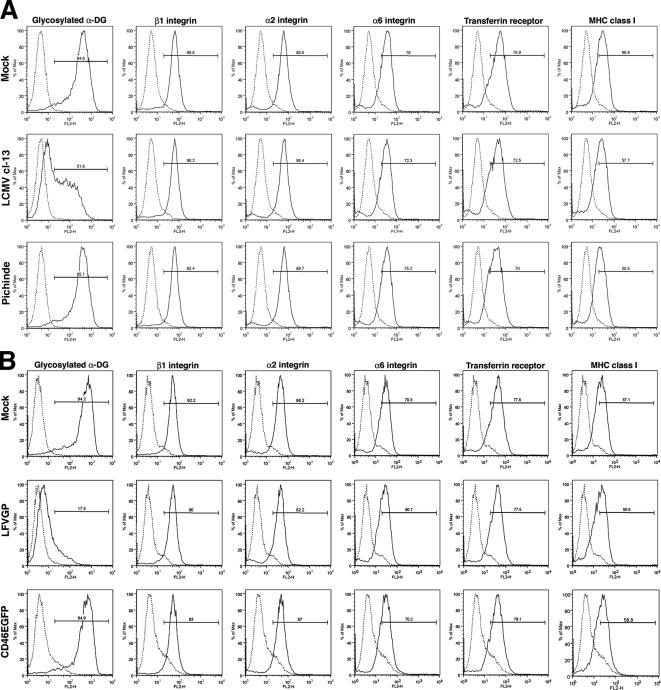
Figure 3.
Virus infection and GP expression do not affect global cell surface expression. HEK293T cells were infected with LCMV cl-13, Pichinde (MOI = 0.1), or mock infected (A), or transfected with empty vector (Mock), LFVGP, and the control protein CD46EGFP (B), which represents a C-terminal fusion of the cellular glycoprotein CD46 with EGFP. After 48 h, the cell surface expression levels of glycosylated α-DG; integrins β1, α2, and α6; transferrin receptor 1; and MHC class I were examined by flow cytometry as described in Materials and Methods. The y-axis represents cell numbers, and the x-axis represents PE fluorescence intensity. Solid line, primary and secondary antibody; broken line, secondary antibody/isotype control.

