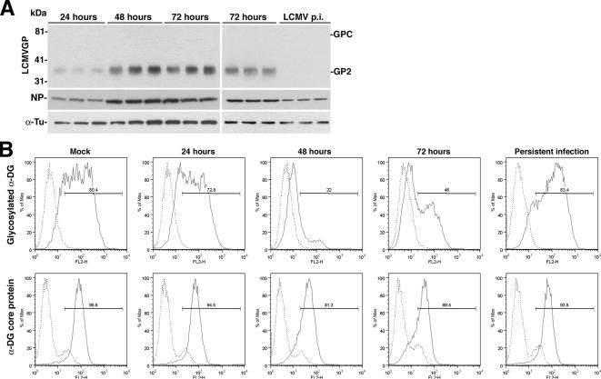
Figure 4.
The virus-induced reduction of functional α-DG expression is reversible. (A) Detection of viral protein-infected cells: triplicate samples of A549 cells infected with LCMV cl-13 for 24–72 h or persistently infected (LCMV p.i.) for 2 wk were lysed, total cell protein was extracted, and cells were probed with mAb 83.6 to GP and polyclonal antibody to NP. For normalization, α-tubulin was detected. The positions of NP, the GPC, and mature GP2 are indicated. (B) Expression of functional α-DG: acutely and persistently infected cells were examined for cell surface expression of glycosylated α-DG and α-DG core protein by flow cytometry as described in Figure 1D. The y-axis represents cell numbers, and the x-axis represents PE fluorescence intensity. Solid line, primary and secondary antibody; broken line, secondary antibody.

