Figure 10.
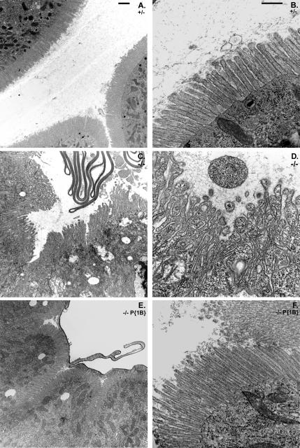
Oral infection with P. entomophila causes a rapid destabilization of the BB in the Myo1B mutant gut. (A) Low-magnification TEM of Myo1B heterozygote 2 h after infection. The BB is intact and cells seems normal. (B) High-magnification TEM of Myo1B heterozygote control 2 h postinfection. (C) Low-magnification TEM of first instar Myo1B mutant gut 2 h after infection with P. entomophila. MV are fused, elongated, and shedding cytoplasm-filled membrane into the lumen. (D) High-magnification TEM of Myo1B mutant midgut 2 h after infection showing severe cytoskeletal damage induced by P. entomophila infection. MV are elongated, fused, and are destabilized. Also note that the MV are filled with cytoplasm, and they are detaching from the enterocyte. (E) Low-magnification TEM of P{Myo1B} rescue gut 2 h after infection. The BB is intact and cells appear normal. (F) High-magnification TEM of P{Myo1B} rescue 2 h post-infection. Bar, 2 μm (A, C, and E) and 500 nm (B, D, and F).

