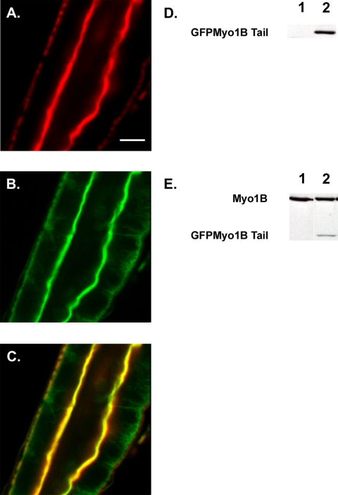
Figure 2.
The tail domain of Myo1B is sufficient for localization. (A) Middle midgut actin visualized with Alexa 568 phalloidin. (B) GFP Myo1B Tail visualized with anti-GFP. (C) Overlay of A and B showing that the tail domain of Myo1B is sufficient for localization to the BB domain of the midgut enterocyte. (D) Western blot probed with anti-GFP to verify expression of GFP Myo1B Tail. Lane 1, WT; lane 2, tail-expressing line. (E) Western blot probed with anti-Myo1B to verify expression of GFP Myo1B tail. Lane 1, WT; lane 2, tail-expressing line. Bar, 20 μm (A–C).

