Figure 4.
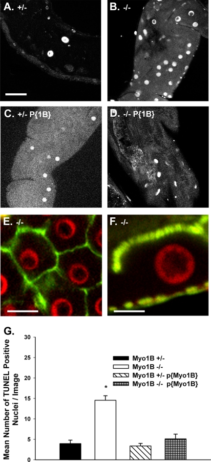
Myo1B mutant larvae have increased numbers of TUNEL-positive nuclei. All TUNEL assay images were taken from same region of the anterior midgut, on the focal plane of the enterocyte nuclei. (A) Myo1B heterozygous control. (B) Myo1B mutant. (C) P{Myo1B} heterozygous (D) P{Myo1B} homozygous. (E) En face confocal image of Myo1B mutant midgut shows that TUNEL-positive nuclei are contained within the junctional boundaries of the midgut enterocytes. (F) Longitudinal section through Myo1B mutant midgut enterocyte. TUNEL staining is specific to enterocyte nuclei. The apical BB domain is at the top of the image, and the gut muscle layer is at the bottom. (G) Quantification of images showed that the loss of Myo1B resulted in a statistically significant increase in the number of TUNEL-positive nuclei per image. Means were compared using one-way ANOVA, p = 0.000. Bonferroni multiple comparison test showed that the Myo1B mutant was significantly different from the other three genotypes. SEM represented by error bars. Bar, 30 μm (A–D), 20 μm (E), and 10 μm (F).

