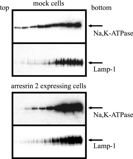
Figure 6.
Cell fractionation of arrestin-expressing and mock-transfected cells by sucrose density gradient centrifugation. COS cells were transfected with vector alone (mock) or arrestin 2. Cells were homogenized in sucrose buffer and centrifuged at 800 × g. Supernatant was layered on a continuous 1.02 to 0.25 M sucrose gradient. After centrifugation, fractions were collected from the top and analyzed by Western blotting with antibodies directed against the Na+,K+-ATPase or the lysosome marker lamp-1. No Na+,K+-ATPase signal was detected in fractions 1 through 11 under either condition. The Western blotting results shown represent fraction numbers 12–20 (from left to right). Typical results from one of four experiments are shown.

